Department of Chemical and Biomolecular Engineering
Feathers of Autumn
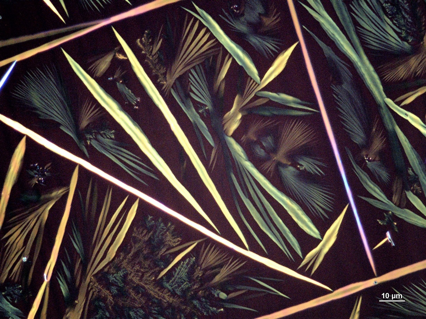
Needles, spherulites, and dendrites are observed in this image of ellipticine, a pharmaceutical used to treat cancer, grown by solution shearing on a polymer substrate. The multitude of crystal morphologies resembles the diversity in color and shape of feathers in autumn. Image coloring slightly enhanced.
Department of Chemical and Biomolecular Engineering, Department of Bioengineering, and Beckman Institute for Advanced Science and Technology
Freeform 3D-Printed Heart Scaffold with Gummy Bear
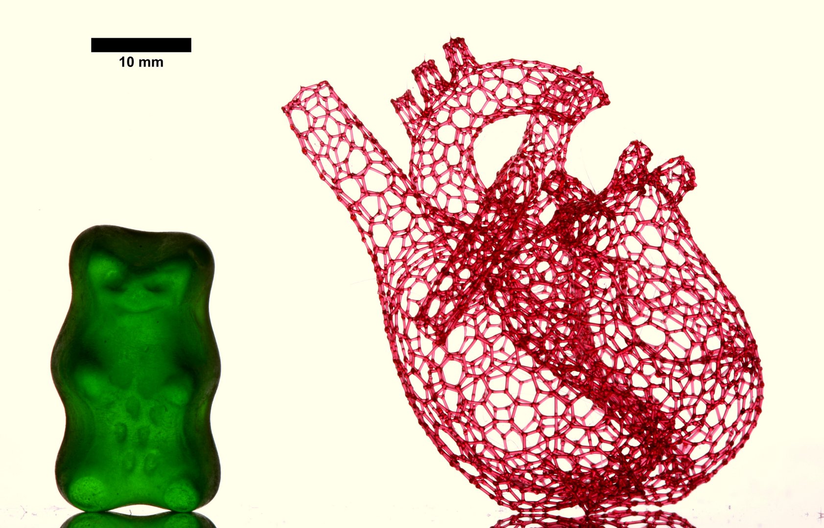
Macro photograph of a vascular template made of a low-calorie sugar substitute. The template is composed of 4,637 filaments, each individually "drawn" by a nozzle translated in 3D space. The template can be coated with a hydrogel then dissolved away, leaving a fully vascularized construct. Gummy bear shown for scale.
Department of Chemistry, Department of Materials Science and Engineering, and Frederick Seitz Materials Research Laboratory
Blooms on Graphene
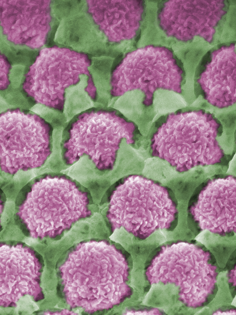
Scanning electron microscope image of a three-dimensionally graphene-sandwiched secondary battery cathode. The fabricated cathode, consisting of an electrically conductive 4~10 layer thick graphene sheet (green color) embedded within electrochemically active vanadium pentoxide (purple color), exhibits a high electrochemical performance including high capacity and long cycling life (>2000 cycles).
Department of Chemistry and the Center for Physics of the Living Cell
A Cell Under Pressure
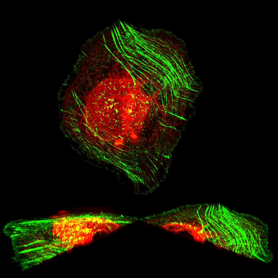
A human osteosarcoma cell shows large deformations on its basal side, a result of rapid exposure to hyper-osmotic pressure. Actin stress fibrils labelled in green maintain the integrity of the cell, but the endoplasmic reticulum and nucleus (in red) are deformed and pushed upwards. Image taken on a Zeiss Elyra SR-SIM scope.
Department of Chemistry
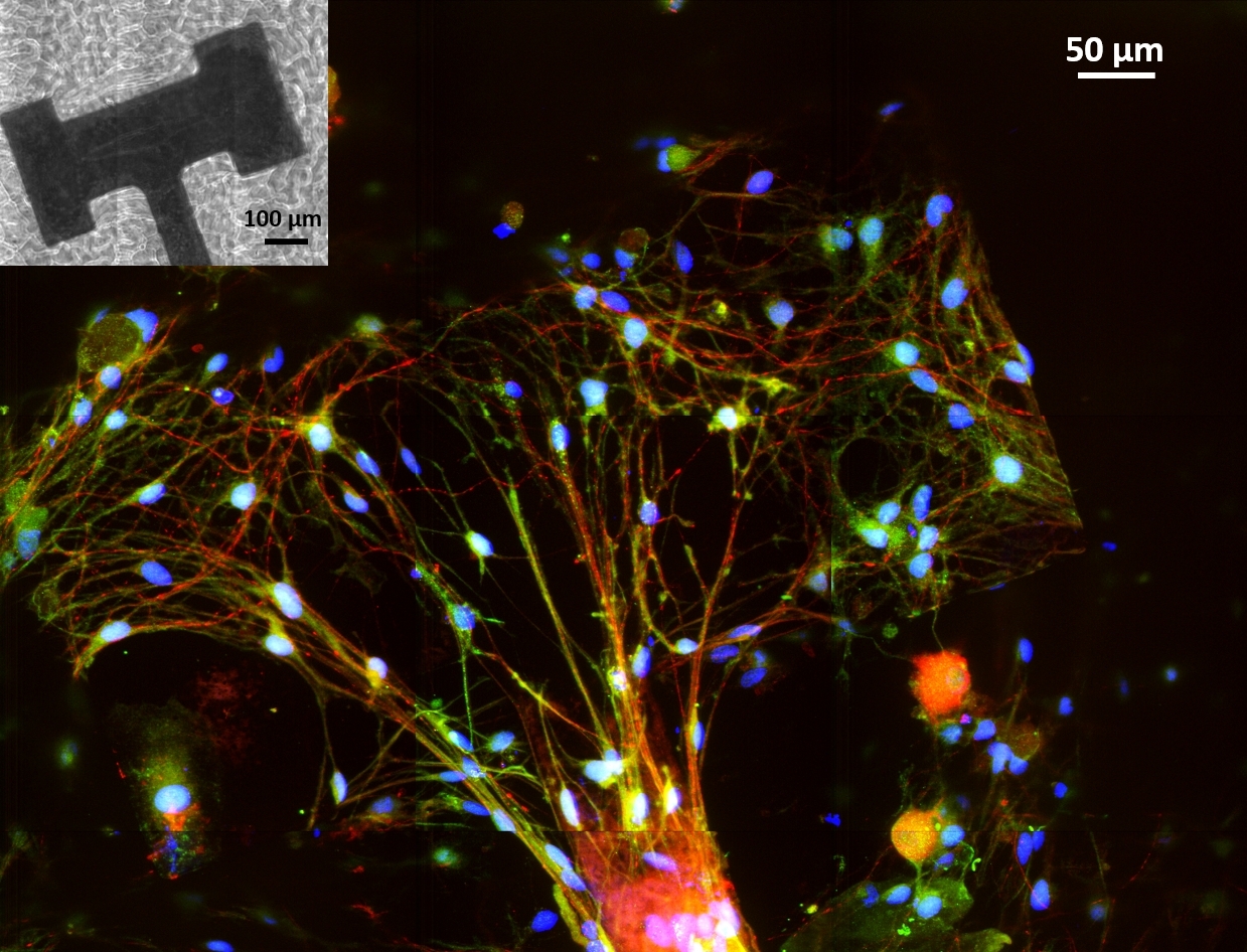
Contact Guidance of Neuronal Network Through Chemistry
Confocal fluorescence micrograph of primary dorsal root ganglion cells cultured on a Silicon 3D scaffold (inset: light micrograph) coated with a custom cell-adhesive protein. The neurons (red) and supporting glial cells (green) form complex intertwining networks with increased time in culture that abide by the geometric cues of the scaffold.